|
|
|
PRODUCTS LIST
Home > Product Show
|
Dual Energy Phantom
Product Numbers: 20206213582
Product description: The phantom is specially designed for dual energy (DE) purposes and can be used for quality assurance, scanner performance and evaluation of different DE post-processing techniques.

|
|
|
INTRODUCTION
Dual Energy Phantom
The phantom is specially designed for dual energy (DE) purposes and can be used for quality assurance, scanner performance and evaluation of different DE post-processing techniques.
Research in computed tomography is currently focused on using Dual Energy (DE) to distinguish between different tissues on CT images.
The DEP-002 is the first phantom providing the opportunity to test CT-scanner performance and to evaluate different DE post processing techniques.
Therefore the phantom provides different virtual non-contrast lesions.
The different cylindrical lesions consists of Ca++ or iodine. For example, in the CT-images some lesion‘s CT-values (HU) can be detected as equal to the surrounding material at one energy (e.g. 120 kV) and with a contrast at other energies (e.g. 80 / 140 kV).
The DEP-002 fits to our additionally available thorax phantom. Extension rings, to simulate obese patients are available, as well.
Specifications
|
| Phantom diameter: |
100 mm |
| Phantom length: |
100 mm |
| Phantom weight: |
0.9 kg |
| Material: |
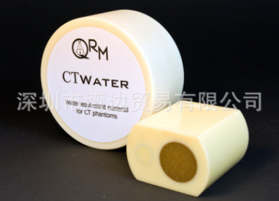
CTwater (0 HU at 80 - 140 kV)
CTiodine (solid iodine)
CaHA (Ca++) |
Specification of lesions
|
| dimension of lesions |
8 x 10 mm cylinder
8 x 5 mm cylinder
1 calibration cylinder 25 mm |
| Layer A: |
Ca++ (200, 400, 600 HU)
Iodine (200, 400, 600 HU |
| Layer B: |
Half cylinder Ca++ (200, 400, 600 HU)
Half cylinder Iodine (200, 400, 600 HU
Full cylinder (25, 50, 100 HU) |
| Layer C: |
Ca++ (-140, -140 HU)
Iodine + Ca++ (0, 0 HU) |
ConeBeam Phantom
The QRM-ConeBeam-Phantom is the most suitable tool to evaluate the imaging performance of cone-beam and flat-panel CT scanners.
These scanners include a wide range technical solutions, e.g. the DVT x-ray scanners, mainly used for 3-D dental imaging, C-arm or angio x-ray machines with options for 3-D imaging or CT scanners with flat-panel detectors covering a large scan volume.
The ConeBeam-Phantom was designed to cover the whole range of image quality achievable with this type of imaging equipment.
The three different low contrast sections provide contrasts between 3 Hounsfield Units and 200 HU to account for the large variation in low contrast capabilities.
Spatial resolution bar patterns range from 4 to 30 lp/cm and an additional edge insert allows to assess the system MTF in different orientations. The image contrast and HU-scale section helps to quantify the machines Hounsfield scale and the bone contrast achieved with different settings.
The ConeBeam Phantom is essential to fully quantify the imaging performance of such scanners and to compare different products or technical solutions.
In addition to the overall image quality of cone- beam scanners, the assiciated dose should be measured using CT dosimetry phantoms (please see our dose phantoms at the dosimetry section). The CT Dose Index (CTDI) methodology can be applied to all types of cone-beamscanners.
Specifications
|
| Overall dimensions: |
| Phantom diameter: |
160 mm |
| Phantom length: |
143 mm |
| Section height: |
20 mm |
Section A, B, C
Contrast resolution: |
| Section A: |
contrast steps: -60, -90, -120, -200 HU |
| Section B: |
contrast steps: -20, -25, -30, -45 HU |
| Section C: |
contrast steps: -3, -5, -10, -15 HU |
| Diameter of Low Contrast Inserts: |
2, 4, 8, 16, 32 mm, respectively |
Section D
Spatial resolution: |
| 14 circular aligned line pattern: |
4 to 30 lp/cm |
Section E
Noise & scaling: |
| 3 inserts D = 24 mm |
bone equivalent (400 mg HA / ccm
water equivalent (CTwater)
air |
Section F
MTF wedges: |
| two perpendicularrly aligned wedges: |
PTFE 20 x 16 x 80 mm |
Section G
Plain: |
| For adapter plate: |
optional custom-made adapter |
Wire MTF Phantom
The Wire MTF Phantom is a perfect tool to assess in-plane spatial resolution of any 3D X-ray imaging system. Different diameters of wire and materials are available.
The QRM Wire Phantom is based on a cylinder containing ONE or more wires in solid material aligned parallel to the phantom axis of rotation.
As variation, one of two wires can be placed slightly off center, the other one in the perifery to allow estimating image quality in plane at two different positions.
Point Spread Function (PSF) and Modulation Transfer Function (MTF) can so easily be investigated.
Different wire diameters are available as well as different materials and positions of the wires inside the phantom.
Specifications
|
| Standard phantom length: |
60 mm |
| Standard phantom diameter: |
45 mm |
| D100 phantom length: |
100 mm |
| D100 phantom diameter: |
100 mm
fits into thorax/abdomen phantom |
| Standard wires: |
tungsten, 50 µm
other diameters and materials upon request |
Spiral / Helical CT 3D Low-Contrast Resolution Phantom
Measure the in-plane and axial low-contrast resolution. Optimize tube current, collimation, pitch and image reconstruction for the desired low-contrast resolution in all types of clinical applications.
The Phantom has been designed to evaluate the imaging capabilities of a CT scanner in the x/y-plane as well as in the axial-plane. The CT’s low-contrast resolution capabilities can be obtained by a single spiral scan using axial images and coronal reformations. The phantom visualizes the impact of all scan, image reconstruction, and display parameters.
Several series of spheres with diameter variing from 3 mm to 8 mm are mounted in a cylindrical body of homogeneous material (resin). Its overall dimensions are chosen such that it fits in our oval Standard Thorax Phantom QRM-Thorax.
The low-contrast inserts are placed in a uniform tissue-equivalent back-ground. In order to mimic imaging conditions in either head or body applications, a contrast of -10HU or -20HU can be ordered. Other contrast levels are available on request.
Evaluation of the 3D Phantom: The scanner’s 3D-visualization software displays saggital (upper left), coronal reformations (upper right), and the original axial image (lower left).
Specifications
|
| Phantom diameter: |
100 mm |
| Phantom length: |
100 mm |
| Phantom weight: |
0.9 kg |
| Material: |
tissue-equivalent plastic, typ. 35 HU (@ 120 kV) |
| Contrast inserts: |
-10 HU or -20 HU relative to background |
| Cylindrical contrast insert: |
diameter 20 mm,
length 25 mm |
| Spherical contrast insert: |
9 spheres diameter 3mm
9 spheres diameter 4mm
9 spheres diameter 5mm
9 spheres diameter 6mm
7 spheres diameter 8mm |
| Accuracy: |
±3% of specified values
±1% of certified values |
|